Home

Monastère accident vasculaire cérébral Le propriétaire cul de sac dural Lune et lautre Particulier Écossais

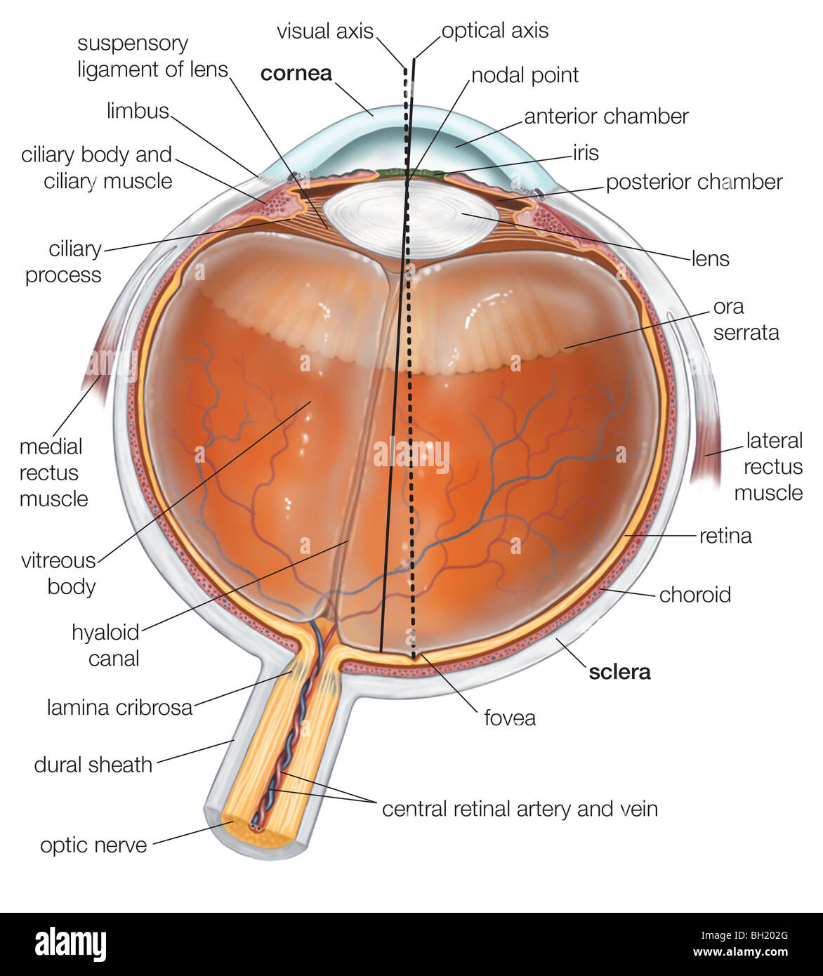
Dural sheath hi-res stock photography and images - Alamy

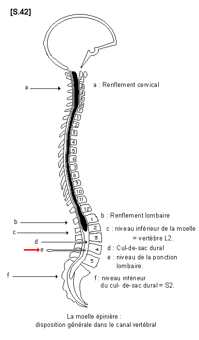
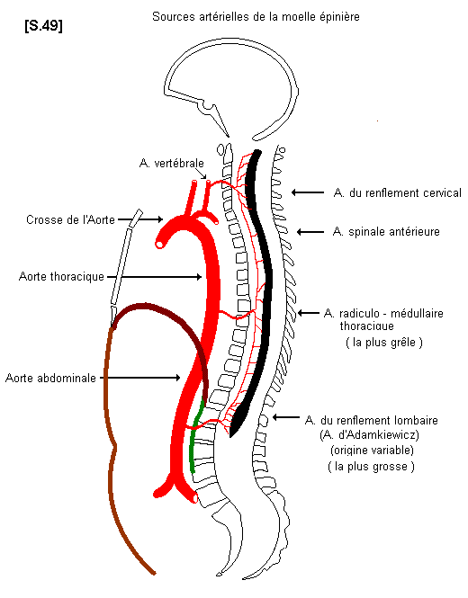
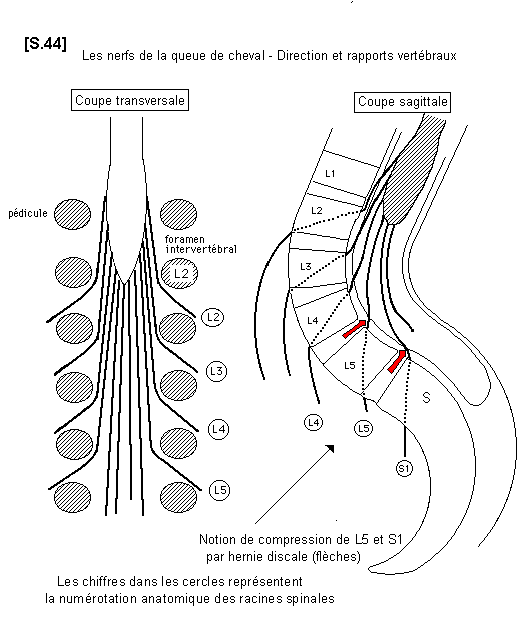
La moelle épinière (1) - Anatomie descriptive

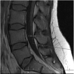
AJNS – African Journal of Neurological Sciences | » TERMINAISON DU CÔNE  MÉDULLAIRE, DU SAC DURAL ET PROFONDEUR DU CANAL VERTÉBRAL CHEZ LE NOIR  AFRICAIN
AJNS – African Journal of Neurological Sciences | » TERMINAISON DU CÔNE MÉDULLAIRE, DU SAC DURAL ET PROFONDEUR DU CANAL VERTÉBRAL CHEZ LE NOIR AFRICAIN

La moelle épinière (1) - Anatomie descriptive

Surgical histopathology of a filar anomaly as an additional tethering  element associated with closed spinal dysraphism of primar
Surgical histopathology of a filar anomaly as an additional tethering element associated with closed spinal dysraphism of primar

Diapositive 1
Diapositive 1

La moelle épinière (1) - Anatomie descriptive

Anatomy of the Spinal Meninges | SpringerLink

a Schematic drawing of the pathophysiology of spinal extradural... |  Download Scientific Diagram
a Schematic drawing of the pathophysiology of spinal extradural... | Download Scientific Diagram

of the Spinal Meninges | Musculoskeletal Key

Syndromes topographiques Dr kohil - YouTube

Spinal cord | Systems | Aula de Anatomia

Dural sac: Anatomy, roles and pathologies | Back pain popularized by health  professionals
Dural sac: Anatomy, roles and pathologies | Back pain popularized by health professionals

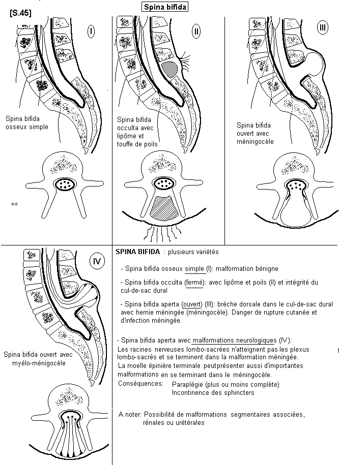
Congenital dermal sinus and filar lipoma located in close proximity at the dural  cul-de-sac mimicking limited dorsal myeloschisis - ScienceDirect
Congenital dermal sinus and filar lipoma located in close proximity at the dural cul-de-sac mimicking limited dorsal myeloschisis - ScienceDirect
Case 2. A 3-month-old girl with caudal type lipoma. (A) Photograph... |  Download Scientific Diagram
Case 2. A 3-month-old girl with caudal type lipoma. (A) Photograph... | Download Scientific Diagram

INSERTIONS DE LA DUREMERE SUR LE RACHIS Une anatomie redécouverte

dure-mère | GDT
dure-mère | GDT

Sac dural : Anatomie, rôles et pathologies | Le mal de dos vulgarisé par  des professionnels de santé
Sac dural : Anatomie, rôles et pathologies | Le mal de dos vulgarisé par des professionnels de santé

Dural sheath hi-res stock photography and images - Alamy

Signals that go with the flow: Trends in Neurosciences

Schematic illustrating the pro-nociceptive mechanism of CatS/FKN... |  Download Scientific Diagram
Schematic illustrating the pro-nociceptive mechanism of CatS/FKN... | Download Scientific Diagram

Diapositive 1
Diapositive 1

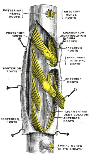
Denticulate ligaments - Wikipedia

Thecal sac - Wikipedia

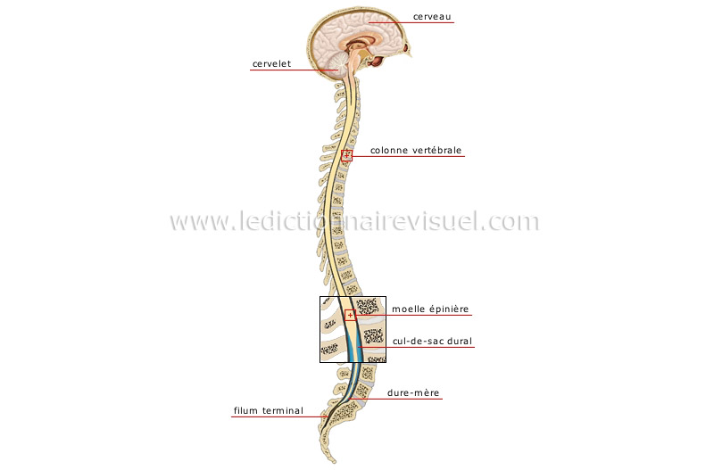
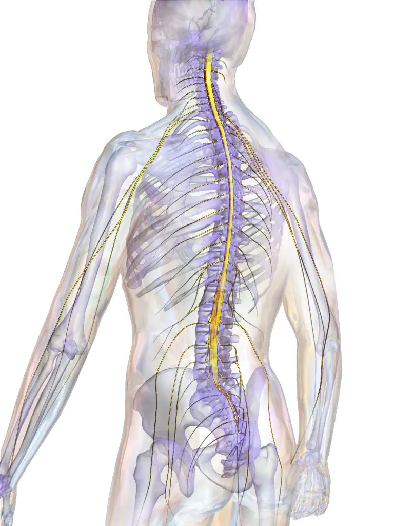
être humain > anatomie > système nerveux > système nerveux central image -  Dictionnaire Visuel
être humain > anatomie > système nerveux > système nerveux central image - Dictionnaire Visuel

La moelle épinière (1) - Anatomie descriptive

Text-book of ophthalmology . e sclera arc reflectedbackwards and form the  dural sheath, 1). which envelops the nerve loosely. Between these  twosheaths lies a third, the arachnoid sheath, A, which divides the
Text-book of ophthalmology . e sclera arc reflectedbackwards and form the dural sheath, 1). which envelops the nerve loosely. Between these twosheaths lies a third, the arachnoid sheath, A, which divides the